
Pregnancy is a voyage that requires love, care, and support for the mother and little one—fetal ultrasounds aid in ensuring that the baby is healthy and safe. Pregnancy scans are performed to receive detailed insights from the womb, which aids healthcare providers in tracking progress, evaluating well-being, and identifying possible complications. Not only this, pregnancy scans facilitate the healthcare providers to detect various chromosomal abnormalities like Down Syndrome, Trisomy-18, spinal issues, and many more. Fetal ultrasounds act as a backbone and the only way out to ensure whether the baby is developing properly or not. The procedure of performing ultrasounds is really easy and risk-free.
What is Fetal Ultrasound?
Ultrasound performed during pregnancy is referred to as fetal ultrasound or prenatal ultrasound. It is a safe imaging method that supports the health of the pregnancy. It is performed for various reasons, such as confirming pregnancy, monitoring the growth of the fetus, and evaluating overall health. Here is an in-depth examination of the pregnancy scans offered to almost every pregnant woman.
A comprehensive range of essential pregnancy scans are offered to pregnant women to check the movement and health of the fetus.

It is one of the early pregnancy scans performed in the early stages to ensure the health and safety of the pregnancy, usually between the 6 to 12 weeks. This ultrasound is performed to confirm the pregnancy in the uterus, determine the number of embryos, estimate the due date, and check the fetus for early abnormalities. Apart from this, this early pregnancy scan is crucial to those women who are experiencing pain or bleeding and have had previous miscarriages or ectopic pregnancies.


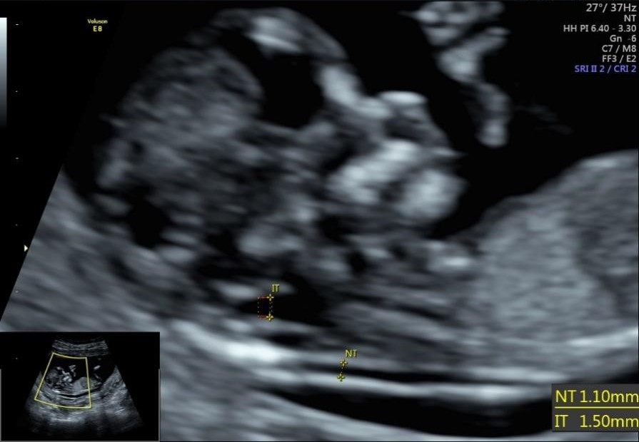
This scan is a non-invasive ultrasound conducted between 11 and 14 weeks of pregnancy to measure the thickness of fluid present at the back of the baby’s neck. The amount of fluid detected behind the neck helps assess the risk of chromosomal abnormalities, neural tube defects, abdominal wall defects, limb abnormalities, and congenital heart disease in the fetus. This essential scan is referred to every expecting woman as it provides an early insight into the baby’s development.


This scan is also referred to as a “Mid-Pregnancy Ultrasound Scan.” This scan is just a screening measure that thoroughly assesses the development and growth of the fetus. It checks for major physical abnormalities in the fetus and provides an early opportunity to detect congenital anomalies. A Fetal Anomaly Scan facilitates timely medical interventions if found necessary.


This pregnancy scan, otherwise called an “Anatomy Scan, " offers a detailed examination of the baby’s anatomy. This scan is used to identify rare conditions and major anatomical abnormalities in the fetus's brain, face, spine, heart, stomach, bowel, kidneys, and limbs. Apart from this, it also screens the position of the placenta and the amount of amniotic fluid. Via this scan, the gender of the baby may also be predicted.


A specialized fetal ultrasound focuses on the baby’s heart and evaluates its structure and function. It is performed between 18 and 24 weeks of pregnancy. This fetal echocardiography is usually carried out to diagnose congenital heart defects and other malformations.


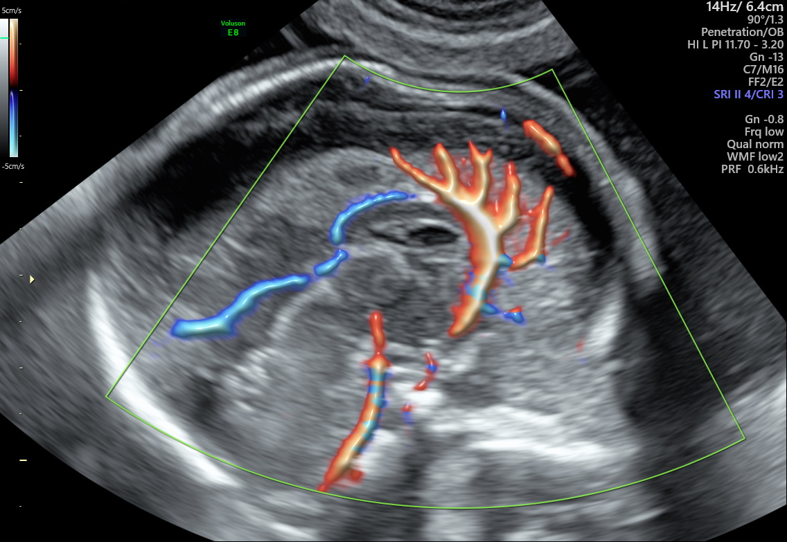
The main purpose of performing this ultrasound is to assess the fetal brain and spinal cord deeply. With this examination, healthcare providers ensure whether the central nervous system is developing correctly and detect other neurological abnormalities.


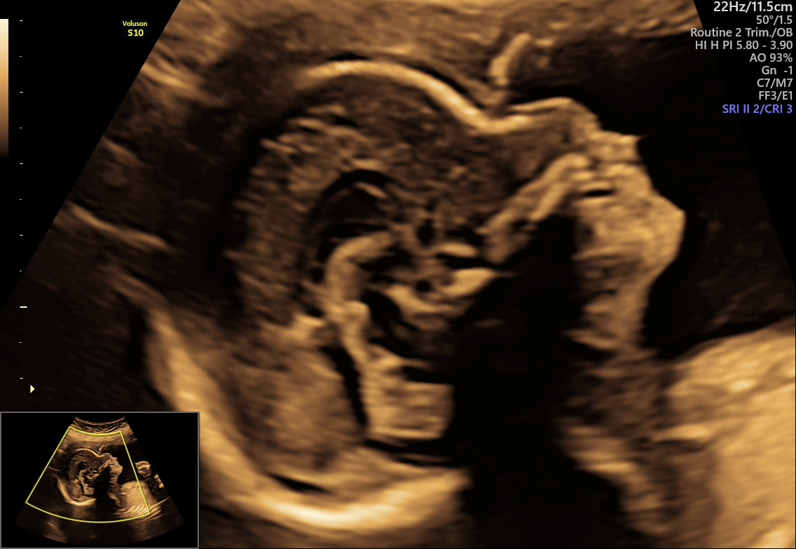
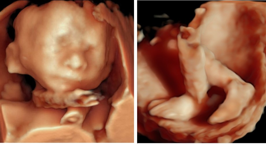
With the 3D pregnancy ultrasound, it has become a lot easier to look at the 3D image of the baby and take a glimpse at the internal organs of the baby. Not only this, these 3D/4D pregnancy scans give a real-time motion of the baby, like kicking and yawning, which enhances the emotional bonding between the baby and parents.


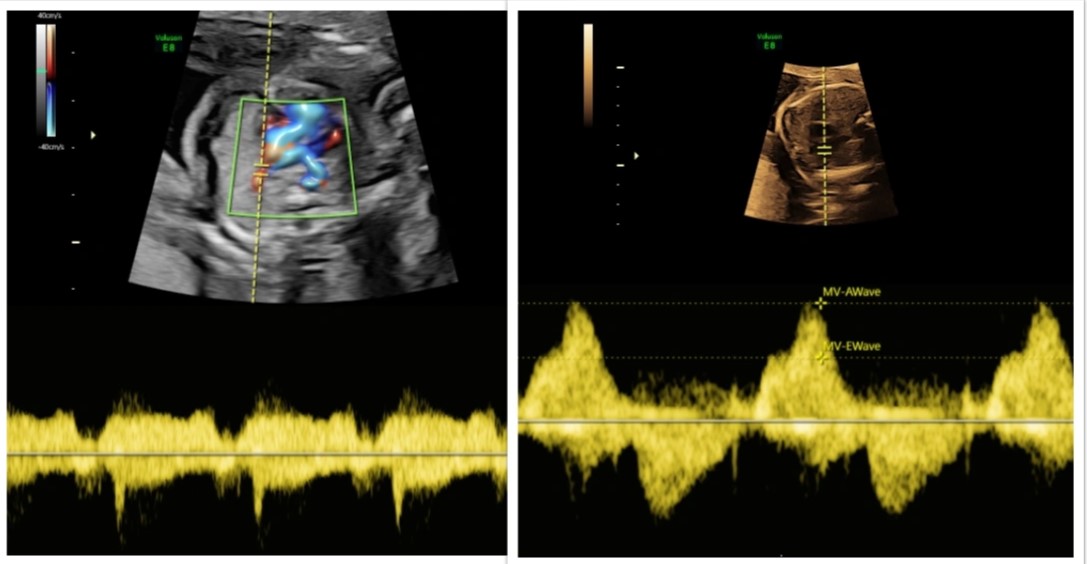
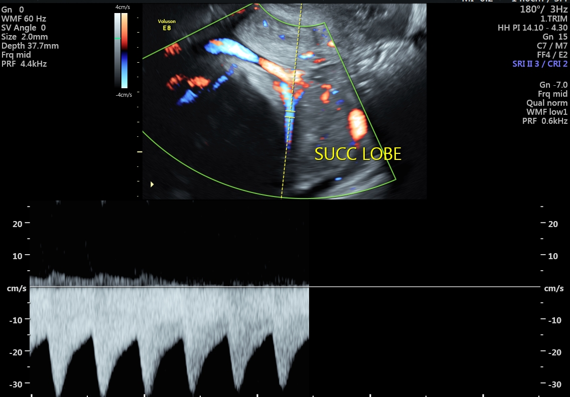
The Fetal Doppler ultrasound keeps track of blood flow in the fetal vessel, which is crucial for managing pregnancies and detecting fetal anemia. A Multivessel Fetal Doppler Monitors Fetal Growth Restriction (FGR). These help in planning proper medical aid and ensure the health and well-being of the fetus throughout the pregnancy.


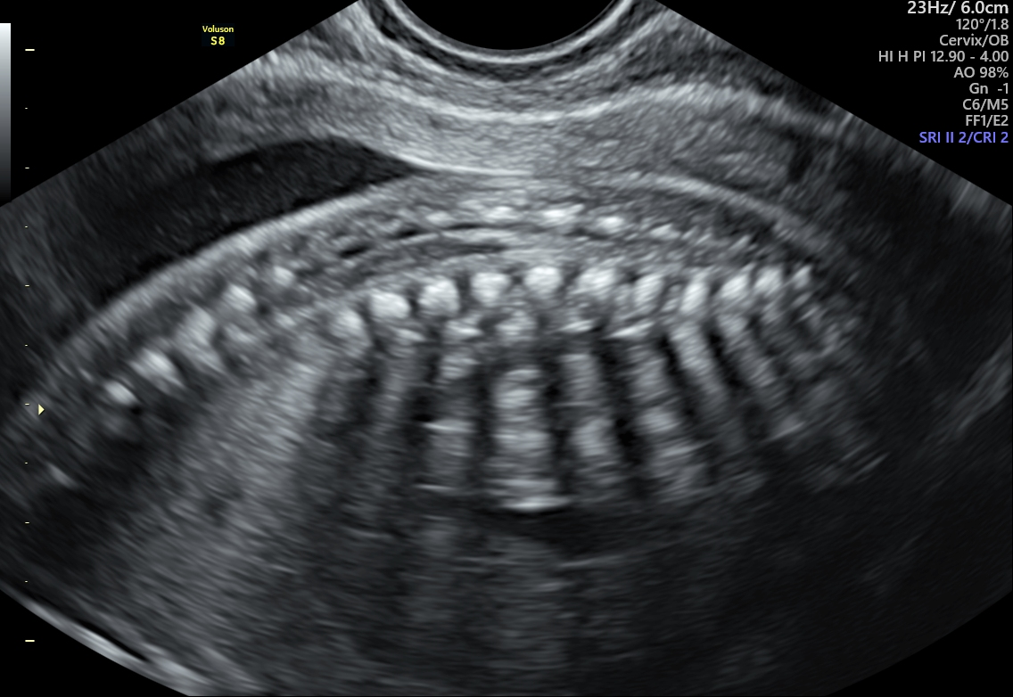
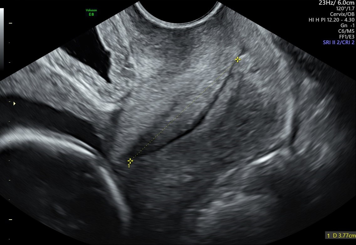
This ultrasound is used to measure cervical length and changes in the cervix. This assessment helps in determining the risk of preterm labor or other conditions that could lead to premature delivery (before 37 weeks of pregnancy). This scan is crucial to identify those women who are at higher risk of delivering their babies early; hence, with timely medical aid, it can be treated.


Specialized scans are required for specific conditions such as fetal growth restriction (FGR) and RH isoimmunization. They help monitor and manage these high-risk pregnancies effectively, providing the necessary information for appropriate medical care.
